
Combat Arts Tricep Injuries Overview
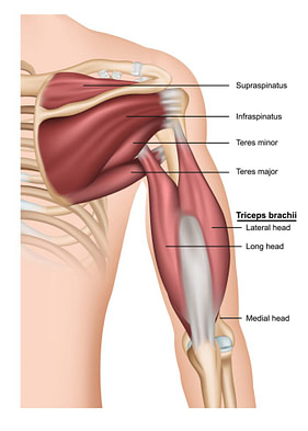
The triceps is the large, thick muscle at the back of the upper arm — the posterior compartment — running from the shoulder blade and the humerus down to the point of the elbow. Its main job is to straighten the elbow: every cross, every push, every post off the mat. A triceps tear that is never treated can leave a visible dent or flattening at the back of the arm, along with lasting weakness extending the elbow, which is why an arm that looks wrong after an injury is worth having examined rather than photographed.
Tricep Injury Causes
Overtraining is the usual background to a triceps tear. Olecranon bursitis sits in the same spot and can muddy the picture, but it is a separate problem rather than a cause of the tear. Increased shoulder flexion stretches the long head of the triceps considerably and raises the load on the tendon. In a martial artist the common mechanism is falling on an outstretched hand: the sudden deceleration piles stress onto a contracted triceps while the elbow is extending. It also happens during dips and bench presses. Striking or jabbing with a posterior elbow into fixed resistance can tear it as well.
Tricep Injury Symptoms
Types of Tricep Injuries
The main injury involving the triceps is a tear. It is uncommon overall, and it is reported more often in people using anabolic steroids, though overuse on its own is enough to do it. Triceps tendinopathy does occur among MMA fighters, but it is not as common as tendinopathy of the knee or shoulder.
Focusing on Arm Strength Can Help Prevent & Manage Tricep Injuries
Subscribe for More Helpful Advice!
Related Injuries
Triceps Tears
The triceps typically have three heads which is how it gets its name. It is located in the dorsal section of the arm. Triceps tears are rarer compared to triceps tendonitis. Having chronic metabolic conditions like diabetes and parathyroid disease increases the chances of triceps tear.
Tricep Tears Symptoms
Source: medicalstocks, Standard License, via Shutterstock
Tricep Tears Causes
A triceps tendon tear or avulsion can take place during a fight on receiving a direct blow to the elbow or falling on an outstretched arm against resistance. Ruptures or avulsions occur at the tendon-bone junction. This due to the eccentric contraction of the muscle. A fighter will experience a painful popping sensation. This is followed by pain and swelling over the posterior elbow. Many fighters will find they cannot extend the elbow against resistance.
To learn how Triceps tears is diagnosed Click Here
Common Tricep Injuries
Other common triceps injuries like tendonitis is fairly common and explained in detail in our Common Injuries section.
Tricep Injury Diagnosis
The diagnosis of triceps injuries tends to be clinical. Plain films will only reveal if there is an underlying fracture. An ultrasound or MRI would be more diagnostic to identify tears in the triceps muscle.
Injury Specific Diagnosis
Triceps Tears
Physical Exam
The physical exam involves isolating the muscle and testing it against resistance. The triceps reflex is tested by tapping the triceps tendon sharply with a reflex hammer, with the shoulder abducted and the elbow flexed to about 90 degrees so the tendon sits under gentle tension — an elbow can be flexed, not abducted. The reflex arc runs through the C7 nerve root, but it needs an intact muscle and tendon to fire: with a complete triceps tendon rupture the reflex is usually reduced or absent, which is a large part of why the test is done.
Imaging
Plain films and MRI capture complete and partial tears of the triceps muscle and tendons. On an X-ray acute tendon tears are seen as bony flecks. This suggests a triceps avulsion injury. It can also be seen on an ultrasound.
For fighters who do not have a complete avulsion, the rupture takes place at the bone-tendon junction and cannot be seen on plain films. Both ultrasound and MRI are used for complete tears and for partial tears that spare the olecranon attachment. In experienced hands ultrasound compares well with MRI for these tears, and either will help locate where the rupture is.
Source: Marc Bruxelle, Standard License, via Shutterstock
Ultrasound is done with the elbow flexed. It shows reduced echogenicity and calcification. MRI can detect abnormalities in tendinopathies. For a snapped triceps, US, MRI, CT, and sonoelastography are all used. Ultrasound is the imaging modality of choice for some as it allows differentiation between tendons and nerves.
Lab Tests
Lab tests are uncommon. The blood tests usually involve looking for metabolic disorders. They include CBC, Hba1c, Urine micro albumin, calcium, phosphorus, PTH, and Vitamin D.
To learn how Triceps tears are treated Click Here
Diagnoses of Common Tricep Injuries
Learn more about how physicians assess injuries through visual inspection, lab tests, x-rays, and other methods in our Common Diagnoses section.
Tricep Injury Treatment
The management of triceps injuries depends on the type of injury. Tendinopathy is managed by settling the pain and rebuilding the tendon’s tolerance to load, starting with isometric holds and progressing gradually under a physiotherapist. Explosive work such as clap push-ups belongs at the far end of that progression, once the tendon handles heavy slow loading without pain — not at the start, and not while it still hurts. For tears, the management is surgical, and depends on the type of tear.


Source: Atstock Productions, Standard License, via Shutterstock
Injury Specific Treatment
Triceps Tears
Emergency
Triceps tears are not medical emergencies. Sometimes, it takes a while for an athlete to identify an injury. If there is no visible deformity, a fighter might disregard the injury as elbow pain. The important step is to identify the injury. Therefore, any pain or symptoms must be referred to an orthopedic doctor right away.
Medical
The treatment for tears is usually surgery. If there is considerable weakness of elbow extension, then surgical repair is advised. For small partial tears, non-operative management has a good prognosis.
After a repair, a brace holding roughly 30 degrees of flexion is typically worn for about 7 weeks, and pushing, resistance work and heavy lifting are avoided for about 12 weeks. Follow the timeline your surgeon gives you rather than this one. Platelet-rich plasma (PRP) injection has been tried for partial tears, but the evidence behind it is limited and it is not an established substitute for surgical repair.
In acute tears of more than 50%, surgical repair is better. Treating chronic ruptures is challenging. Reconstruction with a graft from the Achilles, plantaris, semitendinous, aconeus, latisimus dorsi can be done. Where the triceps are completely avulsed, surgery is advocated. This involves the repair of the avulsed triceps tendon with a Krakow suture pattern to the olecranon. It is done via bone tunnels.
Home
What you do at home after a triceps tear or a repair is set by your surgeon, and it starts with protecting the tendon: the brace, the range limits and the no-lifting window described above come first. Avoid the activity that caused the injury, and anything else that loads the triceps, until you are cleared. The loading work described below belongs to late rehab under a physiotherapist — not to the weeks after a tear or an operation.
Once a clinician clears you to load the tendon, rehab is built on graded resistance — rope triceps press-downs, then single-arm press-downs, adding weight and slowing the lowering phase as tolerance improves. Judge progress on how the tendon responds over the following day or two, and drop back a step if pain climbs. Loading a torn or freshly repaired triceps before that clearance risks re-rupture.
Common Tricep Treatments
Learn more about how healthcare professionals treat injuries using physical therapy, medications, surgery, and other approaches in our Common Treatments section.
