Combat Arts Shoulder Injuries Overview
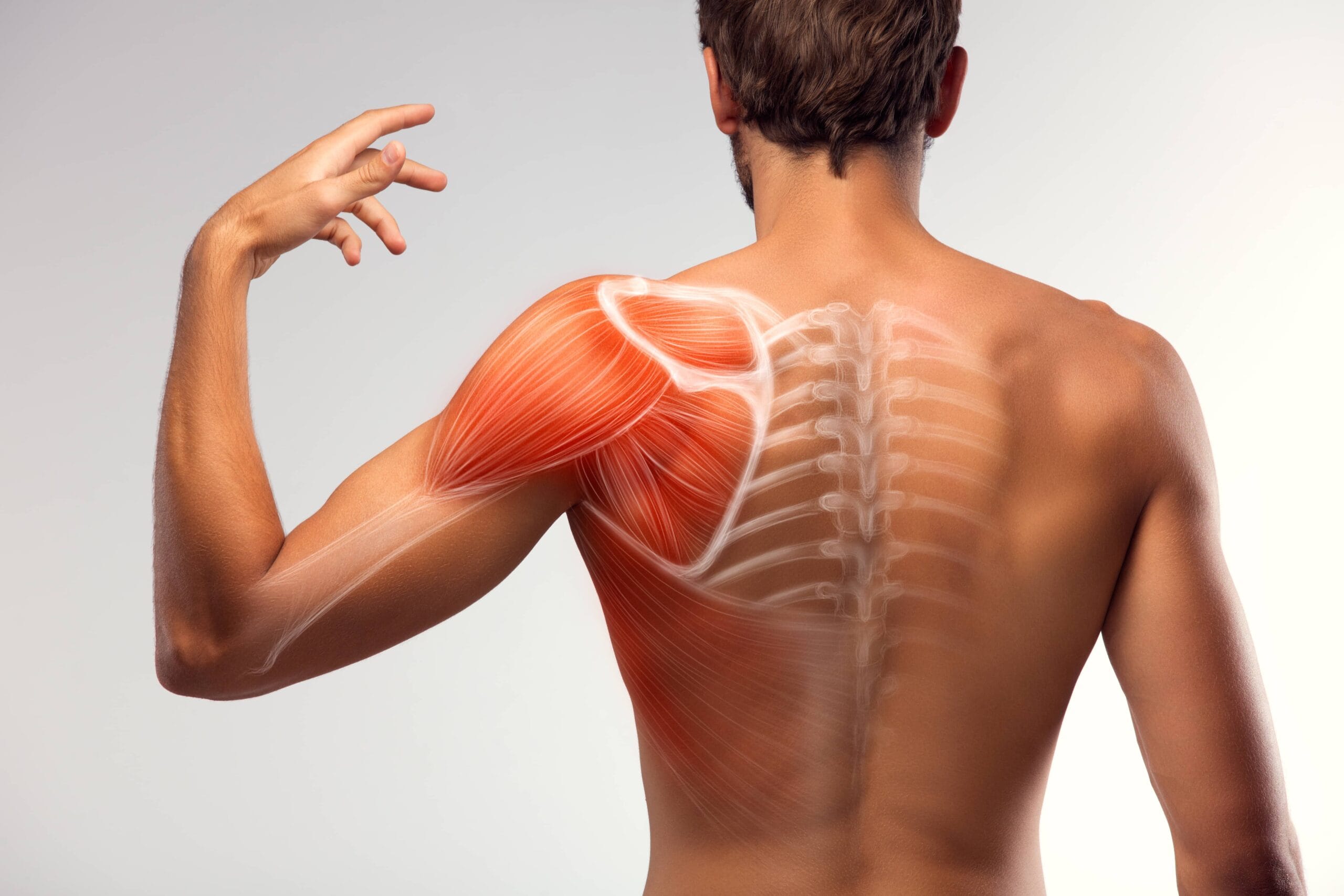
Every time a martial artist performs a throw, punches, or raises the arms, it requires the rotator cuff to engage. Rotator cuff injuries are extremely difficult for martial artists since they weaken the shoulder. Major fight events and heavy sparring often will cause rotator cuff injuries because it is an extremely vulnerable mobile joint that is only held in place by four thick, strong tendons.
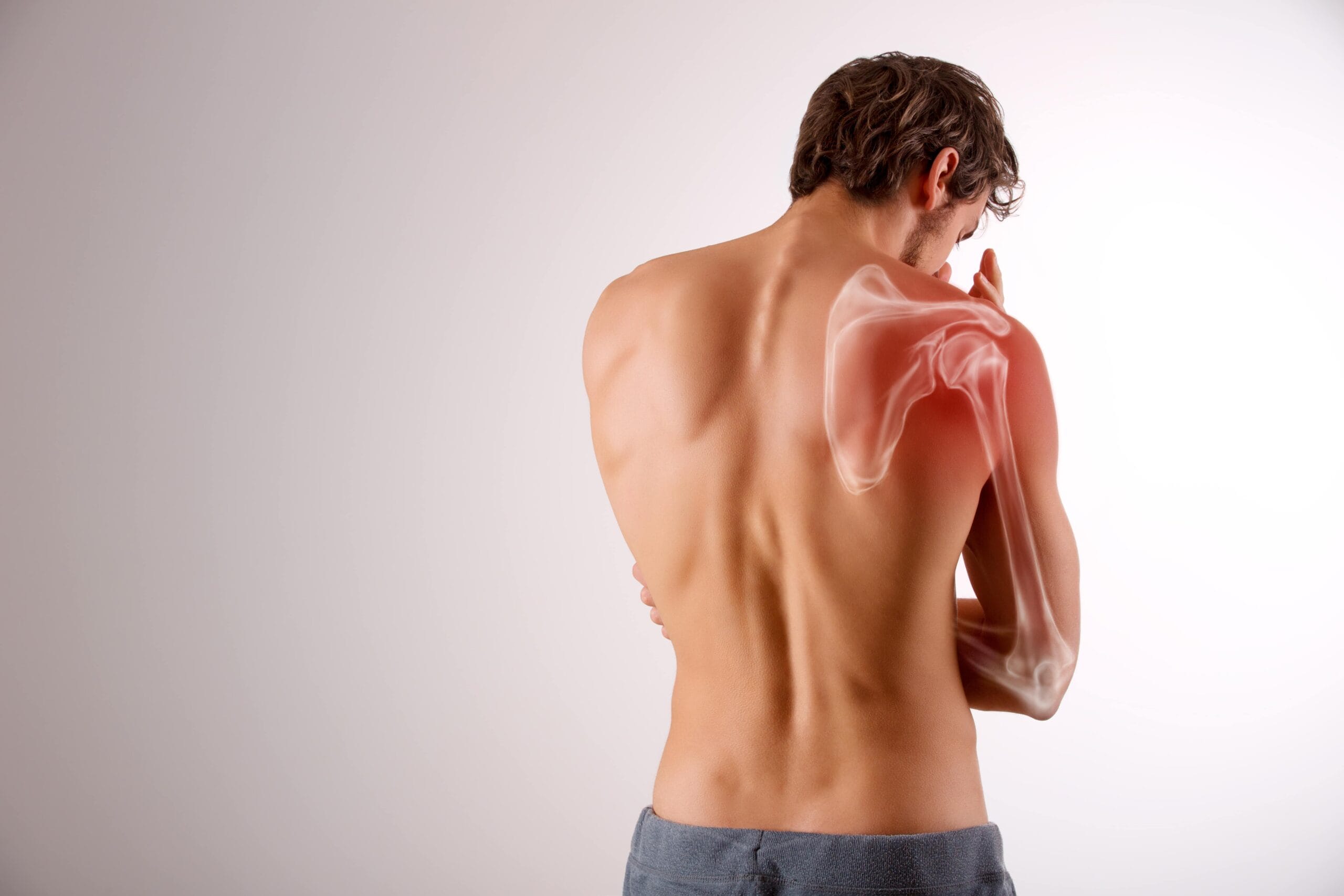
The rotator cuff is a part of the shoulder responsible for securing the upper arm bone or humerus in place while allowing upward movement. It is comprised of three bones that come together: the shoulder blade or scapula, the collarbone or clavicle, and the upper arm bone or humerus.

The rounded ball of the humerus forms a ball-and-socket joint with the shoulder blade. The humerus is held in place by the rotator cuff assembly which is comprised of four tendons that hold the joint together
Shoulder Injury Causes
The shoulder is one of the most mobile joints in the body. MMA fighters typically put their shoulder joints under enormous stress every time they train or fight. Moves like arm bars, Americana, and Kimura exert tremendous pressure on the shoulder. A submission hold, an awkward landing, and forceful contraction all cause the shoulder joint to dislocate and experience tears. It’s for this reason, that the shoulder joint is vulnerable to injury.
There Are Ancient Warrior Ways That Can Help Boost Performance & Recovery
Let Us Teach You!
Shoulder Injury Symptoms

Types of Shoulder Injuries
Injury Specific
A rotator cuff tear or strain is a common injury, especially in combat sports. It may happen suddenly, from a fall or throw, or gradually from repeated stress. Most often the tear or strain will occur in the supraspinatus tendon.
The rotator cuff area is susceptible to bursa damage. The bursae are small lubricating sacs or pillows located between bones that help lubricate and protect against rubbing and grating. These bursa sacs emit protective fluid that ‘oils’ the rotator cuff tendons so they move easily and painlessly. When bursae are damaged they become inflamed or painful. Damage to the rotator cuff may also damage the bursa which intensifies pain and creates additional complications to the injury.
Two Types of Rotator Cuff Tears
Partial Tear. This type of tear is also called an incomplete tear. It damages the tendon but does not completely sever it.
Full Tear. The full tear goes all the way through a tendon or completely severs it from the bone.
Causes
Fraying or wear from repeated stress may be present in a rotator cuff tendon that is then injured during a sudden incident. This fraying may tear unexpectedly during normal activity, such as performing a powerful overhand throw or lifting a heavy object.
Acute Injury: Acute injury tears happen suddenly and usually will cause intense pain. A sense of something snapping in the upper arm or shoulder and immediate weakness or heaviness may be felt. Landing badly from a throw or fall or trying to lift/throw heavy objects can cause this injury. A broken collarbone or dislocated shoulder may create a tear in the rotator cuff tendons.
Degenerative Tear: This type of tear develops slowly, usually from repetitive overuse. It may not cause any pain or other symptoms initially. Once a degenerative tear occurs on one side, one probably exists in the other shoulder, even when no symptoms are present.
Symptoms
These symptoms are indicative of serious rotator cuff issues. It is important to have a torn rotator cuff seen by medical experts. Serious problems may develop over time, such as arthritis or a frozen shoulder.
To learn how Torn or Strained Rotator Cuff is diagnosed, Click Here
50% of all shoulder dislocations are anterior dislocations. That being said, the shoulder can dislocate forward, backward, and downward. It can also be dislocated partially or completely. Extreme rotation can cause it to simply pop out from its socket. Fighters who experience a dislocation, tend to experience a redislocation. This is because the tissue is lax or does not heal properly.
Symptoms
Causes
Anterior dislocations of the shoulder occur due to a blow to an abducted, externally rotated and extended limb. It can also occur when the fighter falls on an outstretched arm due to the force from the posterior humerus.
The arm remains abducted and externally rotated. The acromion looks prominent. Posterior dislocations happen due to a hit to the anterior shoulder. The adducted internally rotated arm bears all the load. The arm is usually in adduction, and internal rotation. Inferiors dislocations occur due to hyperabduction. It can also happen when the abducted arm is overloaded.
To learn how Shoulder Dislocation is diagnosed Click Here
The pectoralis major is a muscle that fans out over the chest and acts as adductor, flexor and internal rotator at the shoulder joint. Tears in this muscle are rare unless, a fighter attempts to lift an opponent or while lifting heavy weights or doing the bench press maneuver. It usually occurs when there’s a violent, eccentric contraction of the muscle.
The pectoralis muscle is a broad, fan-shaped muscle. It has two heads, a sternal head and a clavicular head. The muscle inserts into the humerus. It is responsible for adduction, forward elevation, and internal rotation at the shoulder joint.
Excess tension on a maximally contracted muscle can cause traumatic injury to this muscle. The most common injury is the ‘bench press’ maneuver. Here the arm is abducted and externally rotated. The muscle experiences maximum tension. The injury occurs when the fighter lowers the weight down to the chest. The muscle usually ‘brakes’ the motion. If this eccentric contraction is not smooth due to muscle fatigue, the weight/person slips to one side. It results in a sudden eccentric contraction of the pectoralis major which causes it to rupture.
The muscle also tears when force is applied to a maximally contracted muscle. This happens when a fighter attempts to break a sudden fall in a tackle. It has been observed that direct trauma tears the belly of the muscle. Excess tension causes avulsion of the humeral insertion of the tendon.
To learn how Pectoralis Major Tear are diagnosed, Click Here

Common Shoulder Injuries
Read more about common shoulder injuries like bursitis by visiting our Common Injuries section.
Shoulder Injury Diagnosis
Injuries to the shoulder are mostly diagnosed via physical exam and imaging. Since the shoulder is a cocmplex joint with many muscle, tendon and ligament attachments multiple views and imaging modalities are necessary to evaluate the bone, the cartilage, the muscles and ligaments.
Injury Related Diagnoses
Physical Exam
If degenerative tears or pain develops slowly a medical examination is required to ascertain the level of injury, best treatment, and ensure that there are no other complications such as inflamed bursa or other components such as arthritis or a “pinched nerve.”
Imaging
X-rays do not show the soft tissues of your shoulder such as rotator cuff but are used to assess bone alignment.
Magnetic resonance imaging (MRI) or ultrasound
Shows soft tissue problems such as tears in the rotator cuff tendons. They will help clarify if there is a rotator cuff tear, where it’s located, and the size of the tear. An MRI also shows the quality of the rotator cuff muscles, making it easier to ascertain whether the injury is an "old" one or "new" one.
To learn how Torn or Strained Rotator Cuff is treated, Click Here
Physical Exam
The physical exam is very diagnostic once the range of motion test is done and its observed that the shoulder has a diminished range. In anterior dislocations, the arm is abducted and externally rotated. In thin patients, doctors can palpate a prominent humeral head anteriorly. The posterior void is visible to the naked eye. In posterior dislocations, the arm is internally rotated and adducted. In thin patients, the prominent head is felt posteriorly. Fighters look like they’re guarding the extremity. A detailed neurovascular examination is done before reduction. It is repeated after the reduction as well. In inferior dislocations, the fighter holds his arm above and behind the head. He is unable to adduct the arm.
Imaging
A shoulder x-ray series is more than enough to diagnose a shoulder dislocation. A CT and MRI are required to assess for other fractures. This includes fractures of the glenoid rim or tendon injuries. On a plain film, anterior and inferior dislocations are seen as an incongruence between the humeral head and glenoid outline. If the humeral head is displaced medially and it overlies the glenoid, then it means the dislocation is anterior. Posterior dislocations are difficult to identify on a single AP view film as the congruence is maintained. All dislocations are easily seen on trans-scapular Y views. If bone loss is suspected, then a computed tomography (CT) scan and/or a CT arthrogram is done. Angiography is done if vascular compromise is suspected.
Lab Tests
Lab tests are usually not required for shoulder joint treatment as the management is conservative.
To learn how Shoulder Dislocation is treated, Click Here
During the physical examination, doctors look for swelling and ecchymosis over the anterior chest, the axilla, and the arm. They look for chest asymmetry. Sometimes the muscle belly is prominent or it could be thinned-out. There might be a webbed appearance of the axilla. They will palpate the muscle to see any tenderness over the muscle. Abduction and adduction of the arm are done to 90 degrees to confirm injury to the pectoralis major. Doctors will test the strength of the muscle. The range-of-motion tests will demonstrate any limitation to motion.
X-rays are usually done first. However, they are of limited use. They are done to rule out any bone avulsion. On plain films, the usual shadow of the pectoralis major is lost. The normal anterior axillary fold is also lost.
Ultrasound is more useful due to the low cost and rapid availability. The tears are visible as an uneven echogenicity compared to the contralateral side. Magnetic resonance imaging (MRI) is the preferred imaging modality. This is because it can differentiate between acute or chronic tears. It can also indicate if the tear is complete or partial. MRI is now used to classify the grade and site of injury.
There are four types of injuries. Type I consists of muscle contusions and tears. Type II has partial tears and Type III has complete tears. Type III tears are categorized based on location into A: sternoclavicular, B muscle belly, C myotendinous junction and D insertion.
No lab tests are required for this injury unless surgery is warranted.
To learn how Pectoralis Major Tear are treated, Click Here

Diagnosis of Common Shoulder Injuries
Refer to our Common Diagnoses section about how physicians assess shoulder injuries and test for them.
Shoulder Injury Treatment
The treatment of all shoulder injuries is based on the type of injury. Early identification and diagnosis is key. Often, fighters may choose to sleep off pain in the shoulder joints. Many pectoralis major tears are noticed after four weeks. Most fighters tend to have some tendon degeneration from the repetitive stress which further contributes to the injury. This prolongs and complicates healing.
Injury Specific Treatments
Emergency Treatment
Immediate rest and shoulder sling is the initial step for rotator cuff injuries. Anti-inflammatory medications can reduce the pain. Corticosteroid injections are also given to reduce inflammation. Limit activity and start a shoulder strengthening program if the injury is chronic. For acute injuries, ice the area and seek orthopedic consult.
Medical
A rotator cuff tear may cause further damage and get larger over time. It is very important to seek treatment immediately with an acute injury or if experiencing pain.
Surgery. Research shows surgery is not more effective for the treatment of both full and partial rotator cuff tears and full recovery from rotator cuff injuries can be made using conservative treatment approaches.
Nonsurgical Treatments. Half of all those who get rotator cuff injuries find nonsurgical treatments effectively improve symptoms, including physical therapy, stretches, and exercises designed to increase strength and flexibility of muscles surrounding the shoulder joint.
Therapy device called the ShoulderSphere, which was invented by Dr. Win Chang, MD, strengthens rotator cuff muscles surrounding the shoulder in a functional and multidirectional manner. Win Chang is an orthopedic surgeon and his device is currently used by physical therapy facilities, international sports performance institutions, professional athletes, and more.
Indicators that surgery or nonsurgical methods such as PRP or Stem Cell may be good options:
Home Treatment
Physical therapy that includes core and scapular muscle strengthening are started for those with non-operative management. Subacromial injections with corticosteroids are also given in follow up orthopedic consults.
Emergency Treatment
Shoulder dislocations are usually reduced in the ED. They are not reduced in the ED if the anterior dislocation involved the fracture of humeral neck. This is because it can lead to avascular necrosis
If there have been multiple unsuccessful attempts that could cause include neurovascular compromise, then it is not reduced in the ED.
For posterior dislocations, if the fighter has presented late or more than 6 weeks after the injury, the injury cannot be reduced promptly in the ED. If there are multipart or displaced fractures, then surgical treatment is better. For inferior dislocation, all humeral neck fractures must be reduced via surgery. If vascular injuries are present, surgery is preferred.
Medical
In the ED, there are many techniques to reduce a shoulder dislocation. The first is the scapular manipulation which is about 80% to 100% successful. It can be done in an upright or prone position.
In upright position, the fighter sits up. He can rest the unaffected shoulder against the head of the bed. The doctor stands behind the patient and uses one thumb over the tip of the scapula. He pushes medially while also pushing the acromion inferiorly with the opposite thumb. An assistant can provide traction by holding a fighter’s wrist with one hand. He holds the flexed elbow with other hand and pushes down on the elbow. This is a subtle reduction, though it doesn’t seem so.
The external rotation technique reduces anterior glenohumeral dislocation. This technique overcomes spasm of the humeral internal rotators. It unwinds the joint capsule and enables the external rotators of the rotator cuff. This causes it to pull the humerus posteriorly. It is done with the fighter lying supine. His elbow is flexed to 90 degrees, while he holds the elbow with one hand, and wrist with the other. Gradually, the fighter’s arm is allowed to fall to the side. Every time there is pain, the muscles can relax. Slowly over ten minutes, the arm rotates and reduces without any manipulation.
The Cunningham Technique is where the fighter is seated. The doctor sits in front of the fighter and has the fighter place his hand on top of the doctor’s shoulder. The doctor will rest one arm in patient’s elbow crease and use the opposite hand to massage the biceps, deltoid, and trapezius muscles. The fighter is then asked to pull their shoulder blades together and straighten their posture. This technique is very popular since it doesn’t require any conscious sedation.
The Milch Technique is where the fighter is supine. The doctor places his fingers over the shoulder with thumb in the axilla to stabilize it. The arm is then externally rotated and abducted over head.
The Stimson Technique is where the fighter is prone. His affected arm hangs off the side of bed with 5-15 lbs of weight. In 30 minutes, the dislocation is reduced.
In the traction countertraction technique, a sheet is wrapped under the axilla. Doctors will provide continuous traction at the wrist or elbow. Another doctor provides countertraction with a sheet from the opposite end.
The Spaso Technique is where the fighter lies supine. The doctor grasps his wrist or distal forearm. He lifts it vertically with gentle traction and simultaneous external rotation.
The Fares Technique is where the fighter lies supine with both extremities by their side. The doctor holds the fighters’ wrist and slowly pulls the arm to provide gradual traction. The arm abducts and oscillates back and forth.
The Fulcrum Technique is where the fighter lies supine or sits. A rolled towel is placed in the axilla. The humerus is adducted with concurrent force on the humeral head.
Most of these reductions are done without conscious sedation. Usually, an intraarticular injection of 10 cc of lidocaine or local anesthetic is enough. However, if conscious sedation is required it is done with fentanyl, midazolam, ketamine, propofol or etomidate.
For posterior shoulder reductions, the fighter lies supine. Another doctor applies anterior pressure to the humeral head. Axial traction is applied to the humerus with internal and external rotation.
Home Treatment
After the reduction, the fighter is placed in a sling. A neurovascular exam is done and post-reduction imaging is advised at regular intervals. The fighter must follow-up with an orthopedic surgeon to ensure the reduction is successful.
There’s no emergency treatment except in recognizing that there is a tear in pectoralis major muscle. Since it is a huge muscle, the injury is deceptive. If there is pain, rest, ice, immobilization, and pain medication must be given until consult.
The treatment of a pectoralis major tear depends on how severe the injury is. It also depends on the activity level of the fighter. For complete tears surgery is preferred.
The fighter is put in a sling with the arm adducted and internally rotated. Within two weeks, passive and active range of motion exercises are started. Gradually, return to full range of motion in the next six weeks.
Eight weeks after the injury, progressive resistance exercises can commence. Four months post injury, full resistance training can resume.

Common Treatments
There are various treatments available for shoulder injuries in our Common Treatments section.

