Combat Arts Neck Injuries Overview
Fighters have an extraordinary tolerance to pain. They can power through a move, a grueling workout, or a fight and roll with the punches without batting an eyelid. MMA referee and BJJ black belt John McCarthy knows about it first-hand.
He wasn’t even fighting but teaching when he experienced a neck injury that changed his life forever. While demonstrating how to put a proper d’arce choke, he allowed a student to put one on him. The student did everything right. The move lasted ninety seconds. The following morning, he had numbness and burning right below his left shoulder all the way to his arms. McCarthy had destroyed the discs and the nerves supplying the cervical vertebrae that had already been under strain from years of training.

Mixed martial arts can strain your neck muscles and the cervical spine. And this is even though many fighters do neck strengthening exercises. These exercises can reduce the amount of energy transferred but over time, the sustained injury can affect the vertebrae, the muscles, and ligaments that supply them.
Neck Injury Causes
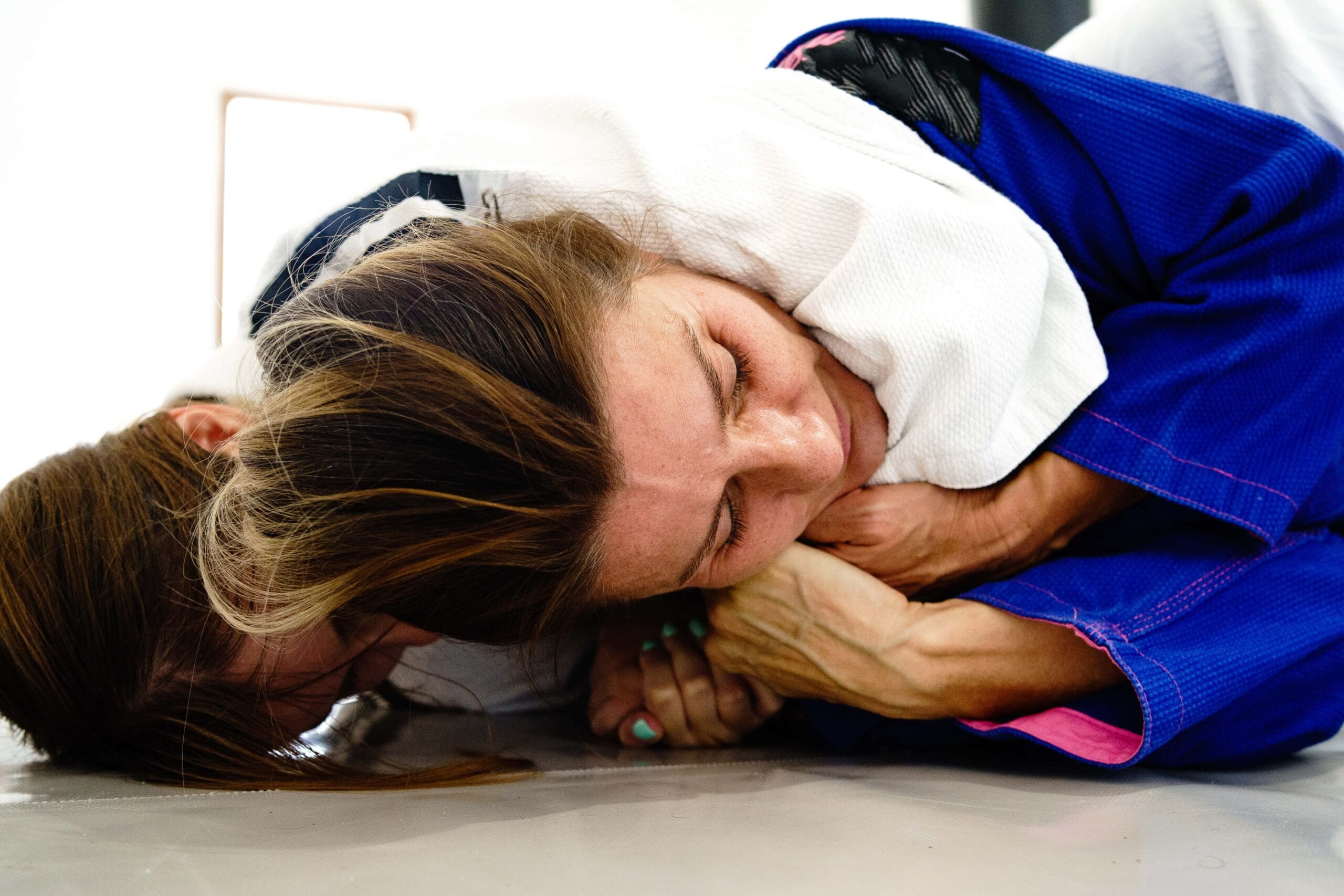
Several moves in mixed martial arts put the cervical spine at risk. In judo, the o-goshi or hip toss is a simple and common maneuver. The fighter will step into the clinch and will swing the opponent over his hips. The opponent will fall on his back. The suplex move in jujitsu, the fighter grabs the opponent by his waist.
With their combined center of gravity moving, the fighter will fall on his back to the mat maintaining his hold on the opponent. A variant of this move, the souplesse, is where the fighter lifts the opponent by the waist and throws him over his shoulder. Before the throw, the opponent is rotated over the upper chest and slammed down. Then there are the chokeholds. A fighter will reach around the neck of an opponent with one hand and complete the choke with the other hand.
All these moves cause force displacement of the head, hyperextension of the neck ligaments, strong flexion of the cervical spine and it’s junction with the occiput. The constant acceleration, sudden deceleration, and biomechanical forces don’t allow the muscles and joints sufficient time to prepare for impact. Therefore, the kinematics of these moves can affect the structures in the neck causing injuries that could be life-altering.
Neck Injury Symptoms

Types of Neck Injuries
Injuries in the neck region are more than simply a pain in the neck. These injuries can involve the bones and joints of the cervical spine, the intervertebral discs that act as shock absorbers between the discs, and the muscles and ligaments that hold them all together.
Got a Pain in the Neck?
Learn Home Remedies Tips and Tricks to Recover Faster
Related Injuries
This neck injury is due to forceful, rapid back-and-forth movement of the neck. It is similar to the cracking of a whip and hence the name. It may involve sprains or strains, injured nerve roots, intervertebral joint stress, and/or hyperextended cervical muscles, ligaments, or discs.
Causes
Whiplash damage may occur during MMA and UFC chokes, uppercuts, throws, or incorrect landings.
Symptoms
Whiplash symptoms may be experienced immediately, or it may be hours or even days before they appear. 50% of athletes can present with whiplash-associated disorders months to years after the initial injury.
To learn how Whiplash is diagnosed, click here.
Overview
In mixed martial arts, the neck encounters forces which cause the neck to be strained. Headgear can lessen the impact of the forces due to linear acceleration. However, most martial arts don’t use any headgear at all. A mat may decrease the impact but the forces acting on the cervical spine and the velocity tends to strain these cervical spinal ligaments and the muscles.
Most cervical sprains and strains are self-limiting. However, it's worth noting that the repeated cervical sprain and strains can further deteriorate the stability of the spine and the integrity of the intervertebral discs.
Causes
There are multiple causes of cervical sprains and strains. A cervical sprain is an injury to the ligaments. These are the connective tissue that holds the vertebrae in place. Cervical strains are injuries to the muscles that support the cervical spine. The cervical spine has to stretch considerably to resist flexion, side flexion and hyperextension.
Several muscles must contract isometrically to maintain the neck’s position. Muscles must also contract eccentrically to counter the forced displacement of the head by an opponent. In addition to all these takedowns and grappling, the environment also poses a threat to the neck. A study showed that 63% of athletes had sustained injuries from impact to the ground or cage.
Symptoms
To learn how Cervical Sprain/strain is diagnosed, click here.
The cervical spine is most susceptible to injury. This is because it highly mobile and the vertebral bodies that make up the cervical spine are small. It supports a heavy head and, in most cases, acts as a lever. The most dislocations and fractures involve the C2 spine in 30% of cases and C7 spine in 27% of cases.
Fractures of the transverse foramen of the vertebral bodies are associated with a blunt cerebrovascular injury. A fighter can have a dislocation where the ligament is torn or fractured or both.
There are four major mechanisms of actions that are associated with various fracture patterns. These are shearing, rotational, flexion, and extension forces. Flexion is associated with bilateral facet dislocation, anterior wedge fracture, flexion teardrop fracture, clay-shoveler fracture, anterior subluxation, and anterior atlantoaxial subluxation.
An extension is associated with posterior atlantoaxial subluxation, hangman fracture, and C1 arch fracture. Lateral flexion causes unilateral occipital condyle fracture. Flexion rotation causes rotatory atlantoaxial dislocation. Extension rotation is associated with articular pillar fracture. Axial loading is seen causing burst fractures or Jefferson fractures. Shearing forces can cause more complex fractures such as odontoid process fractures and occiput condylar fractures.
Fractures are classified according to location or the fracture pattern. Since the anatomy of the spine is unique, cervical injuries are classified occipital-cervical (occiput-C2) and subaxial cervical spine (C3-C7) injuries. Atlas fractures, axis fractures, odontoid fractures fall under the occiput-C2 category. Facet fractures, teardrop fractures, and burst fractures come under C3-C7 fractures.
The fractures may also cause the underlying blood vessels and nerves to be injured. This is what results in the injury to the spinal cord and leads to neurological deficits. In some cases, this deficit can be irreversible.
Learn how Dislocation of the Spine is diagnosed, click here.
A veteran of Brazilian jiu-jitsu, Ricardo Liberio has seven herniated discs. He cannot move without pain anymore but he still rolls. A herniated disc is when the spongy cushion that acts as a shock absorber between your spine, gets compressed, damaged, or starts spilling out into the bony skeleton. A slipped disc is also called a ruptured disc.
A bulging disc is when the ring or annulus that protects the nucleus is intact but protrudes and compresses the nerves. Sometimes, the herniation is severe enough for a free fragment to break through into the spinal canal. Depending on which disc is injured the spine symptoms for a herniated disc differ.
With age, our discs lose their elasticity. They become shorter, less elastic, and less effective as shock absorbers. Repeated trauma can also cause a disc to rupture even if the pressure is slight.
The tough fibrous outer wall or annulus degenerates with time. This degeneration is faster with multiple trauma. If that is coupled with smoking, bad genetics, and occupational hazards, the chance of disc degeneration is much higher. Only 8% of all herniated discs occur in the cervical spine area.
To learn how Ruptured Disc is diagnosed, click here.
Common Injuries
Visit our Common Injuries section to learn more about tendinopathies and how they should be managed.
Neck Injury Diagnosis
The diagnosis of spine injuries requires imaging but also a good physical examination. It’s important to distinguish spine injuries from head injuries. Both require immediate testing and treatment. While most aches and pains are self-limiting, neurological deficits point to a more serious underlying pathology.
If fighters sustain neck injuries, they shouldn’t simply power on. Instead, they must test motor functions in all body parts and then seek medical attention. There may be a delay in symptoms and in case of neurological symptoms, the absence of sensation might flag one’s attention rather than their presence.

While most aches and pains are self-limiting, neurological deficits point to a more serious underlying pathology. If fighters sustain neck injuries, they shouldn’t simply power on. Instead, they must test motor functions in all body parts and then seek medical attention.
There may be a delay in symptoms and in case of neurological symptoms, the absence of sensation might flag one’s attention rather than their presence.
Injury Specific Diagnosis
Physical Exam
A physical exam is important to determine the extent of a whiplash injury. The doctor will test the range of motion of the neck and shoulder. These tests ask fighters to turn from side to side, rotate chin to shoulder, and other flexion and extension movements. Trigger points are assessed. The sensorimotor function is tested and deep tendon reflexes are also tested to rule out any neurological deficit.
Imaging:
A physical exam is important to determine the extent of a whiplash injury.
A CT scan or MRI may be required to ascertain the severity of the injury since extreme whiplash may cause spinal damage to the adjacent bones or discs. MRIs are more sensitive for evaluating the surrounding soft tissue in the cervical area.
Lab Tests
For fighters with whiplash-associated disorders (WAD) to have a few basic blood tests. The complete blood count to check the presence of any infection.
The erythrocyte sedimentation rate (ESR) is done to determine any increased inflammation in the body. The C reactive protein CRP has been a god marker of inflammation to identify injury to muscles and is also done in WAD.
To learn how Whiplash is treated, click here.
Physical Exam
A complete physical examination is done to test all neurological functions. The range of motion of the neck is tested along with deep tendon reflexes. The doctor will also check for point tenderness.
Imaging:
Cervical sprains and strains do not need diagnostic imaging. However, Xrays are ordered just to make sure there’s no underlying fracture. If Xrays prove inconclusive or there is a finding that needs to be seen clearly a CT or MRI is done.
Lab Tests
Blood tests are usually unnecessary for cervical sprains or strains. Inflammatory markers such as CRP and ESR are done if the condition does not resolve in a few days.
To learn how Cervical Sprain or Strain is treated, click here.
The doctor will perform a restricted but thorough neurological exam. This includes testing cranial nerves, sensory function, motor, function, coordination, and reflexes. The respiratory and bowel bladder will also be examined to check for spinal never integrity.
Xrays are the first step to visualize a spine fracture. A CT then provides more detail about the fracture and is usually recommended. CT is the preferred method to examine cervical spine fractures. CT can visualize the shape and size of the canal. The content of the spinal canal and the structures around it are also visible on a CT. The MRI can amplify the nerve roots and any degeneration which is why it’s also helpful to do it.
If immediate surgery is advised, the blood tests are required. These include the CBC, BMP, PT, PTT, ESR, CRP, HbA1c, and urinalysis.
To learn how Dislocation or Fracture of the Spine is treated, click here.
On physical exam, the fighter may or may not have any neurological deficits. However, a neurological exam is important. The doctor will look for signs of numbness and weakness. The fighter is asked to walk normally and on tiptoes. Muscle strength and reflexes are tested. Range of motion tests are done to test flexibility and movement in the joints.
For a ruptured disc, a series of tests are done. Xrays are usually done in the beginning not to confirm or diagnose a herniated disc but to rule out other problems like a fracture, presence of a tumor, or secondary causes of spinal syndromes. MRI provides the most accurate assessment of the cervical spine.
It provides a detailed view of which spine is affected, where the herniation has occurred, and which nerves are affected. An MRI can demonstrate how the nerve has impinged. A CT is not as accurate as an MRI and is usually not helpful for herniated discs unless there’s a reason an MRI cannot be ordered.
A CT myelogram can also demonstrate the size and location of the herniated disc. However, it is invasive. A contrast dye is injected in the spinal fluid and a CT is done. An electromyograph can also show which nerve root is affected by a herniated disc. It’s a helpful test to differentiate between nerve degeneration and nerve root compression.
Blood tests are usually not required to diagnose a herniated disc. If a person is scheduled for surgery then pre-surgical lists of tests are done.
To learn how Ruptured Disc is treated, click here.
Common Diagnoses
In our Common Diagnoses section, read how physicians examine, test and treat common neck injuries.
Neck Injury Treatment
The treatment of cervical injuries runs the gamut from ice to surgery and physical therapy. It depends on the type and severity of the injury. Cervical spinal injuries that cause difficulty of breathing or paralysis are worse and need an immediate surgical intervention when possible.
Associated dislocations and disabilities also affect the treatment. The timing of the intervention is also subject to the type of injury and the symptoms. The medical team will decide the type and timing of the intervention after considering all of the above.

Injury Specific Treatment
Emergency
Apply ice to acute injury. Administer pain medication and monitor. Align head and neck with a small pillow.
Medical
A cervical collar is applied to make sure the cervical spine is stable. Muscle relaxants and physical therapy are started.
Home
Heat or cold can be applied to the area of pain every fifteen minutes. Start stretching and movement exercises to restore normal range of motion of the cervical spine. These exercises include rolling head from side to side and rotating neck in both directions. Physical therapy is prescribed if at home exercises do not relieve any pain. This will strengthen your muscles and also restore motion. Foam collars for 72 hours are helpful but experts warn against using them for longer periods.
Emergency
Wear a soft collar or brace to relieve any neck pain or pressure. Apply an ice pack for 15-30 minutes a day, several times a day to reduce inflammation. While moist heat does loosen up muscles be careful and restrict its use after the initial injury.
Medical
Pain medications like aspirin and ibuprofen can help relieve inflammation and reduce pain. Muscle relaxants can relieve spasms. With the supervision of a medical doctor, massage therapy and ultrasound therapy is started. Some may even recommend, cervical neck traction or isometric exercise.
Home
Rest and ice should enough. Cervical sprains and strains usually resolve in 2- 4 weeks. Until the range of motion and pain is relieved, return to play is not recommended as this would further strain the muscles and ligaments. Severe injuries will take longer to heal.
This is a true medical emergency. Do not move the fighter. Keep them still. Place rolled towels on both sides of the neck and avoid moving this area. Always assume the person has a spinal injury if they had a head-on collision, they complain of severe neck pain and weakness or paralysis. Do not remove any headgear.
Medical treatment can be conservative or surgical. C1-C2 dislocations should always be treated surgically. Initial treatment almost always involves skeletal traction and closed reduction. This includes metal pains places in the skull with a pulley and a rope attached. Many cervical orthoses are used for stabilizing the cervical spine. These can be soft collars or halo vest mobilizations.
Surgery usually involves cervical fusion and instrumentation. Surgical decompression is another alternative. There are various guidelines for surgery of the cervical spine. Atlanto-Occipital Dislocation (AOD) is reduced with positioning, halo vest. Almost all require posterior spinal fusion (PSF) surgery. Occipital Condyle Fractures require Cervical orthosis for 8 weeks in Type I and II while Type III requires PSF.
Atlanto-Axial (C1-C2) Instability (TAL Insufficiency) needs surgery of > 7mm of instability since the spinal cord is at risk. Atlantoaxial Rotary Subluxation is reduced with halo traction. Atlas (C1) Fracture is nonoperative. Cervical orthoses are used here. Traumatic Spondylolisthesis of the Axis (C2) is reduced via surgery if they are severe.
Subaxial Fractures (C3-C7) are treated with a closed reduction if there’s no spinal canal compromise. Cervical orthoses will suffice. For unstable C33-C7 fractures, open reduction with surgery is advised.
Treatment depends on the type and location of the fracture. It also depends on how severe the fracture is and how much displacement has occurred. The presence of spinal cord/nerve compression will also affect the specific therapy. If neurologic dysfunction or spinal cord injury persists then the treatment is more aggressive. Also, the fighters’ age, medical condition, and associated injuries will influence the type of treatment.
Home care for spinal injuries is difficult depending on the type of injury. Most often fighters are kept in traction at the hospital for months especially if there are severe neurological complications. After surgery, complete bed rest is advised.
Ice the area every 15 minutes till pain resolves. And seek medical attention if neurological deficit observed. Do not use any heat or heating pads.
Pain medication is started to reduce inflammation and decrease pain. Muscle relaxants can ease muscle spasms in the cervical area. Spinal injections like epidural or nerve block injections where cortisone or other steroids can help reduce inflammation and improve movement.
For fighters who do not improve with conservative treatment or in those whose bowel and bladder function is compromised surgery may be helpful. The most common surgery for the cervical spine is microdiscectomy, laminectomy, or foraminotomy. In microdiscectomy, an operating microscope is used to remove the fragments of a herniated disc. In a laminectomy, the lamina of the spinal cord is removed to relieve the pressure on the disc. This eases pressure on the nerves and spinal cord. It takes about six to eight weeks to completely recover from this surgery.
If these do not work, then surgeons may go back and perform a spinal fusion after removing the herniated material. They may also replace the disc with artificial disc material. This preserves the motion of the spine while fusion does not allow any further motion once the vertebra is fused.
Physical therapy is advised for those with ruptured discs. This helps strengthen and stretch the muscles of the neck, shoulder, and arms. Alternative therapy includes acupuncture, acupressure, and biofeedback.
Common Treatments
Refer to the Common Treatments section to find out how physical therapy and medications can help neck injuries.

