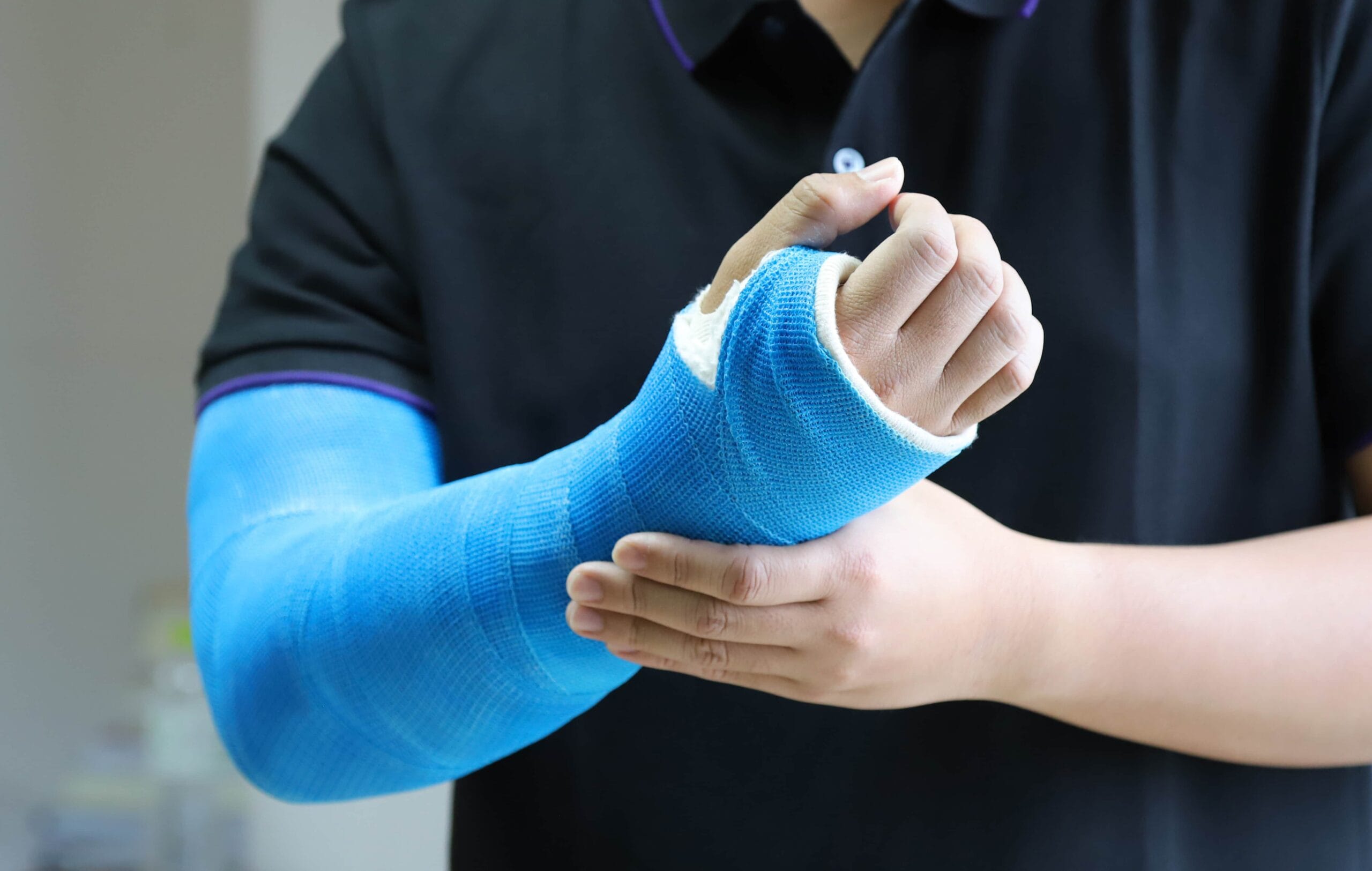
Combat Arts Forearm Injuries Overview
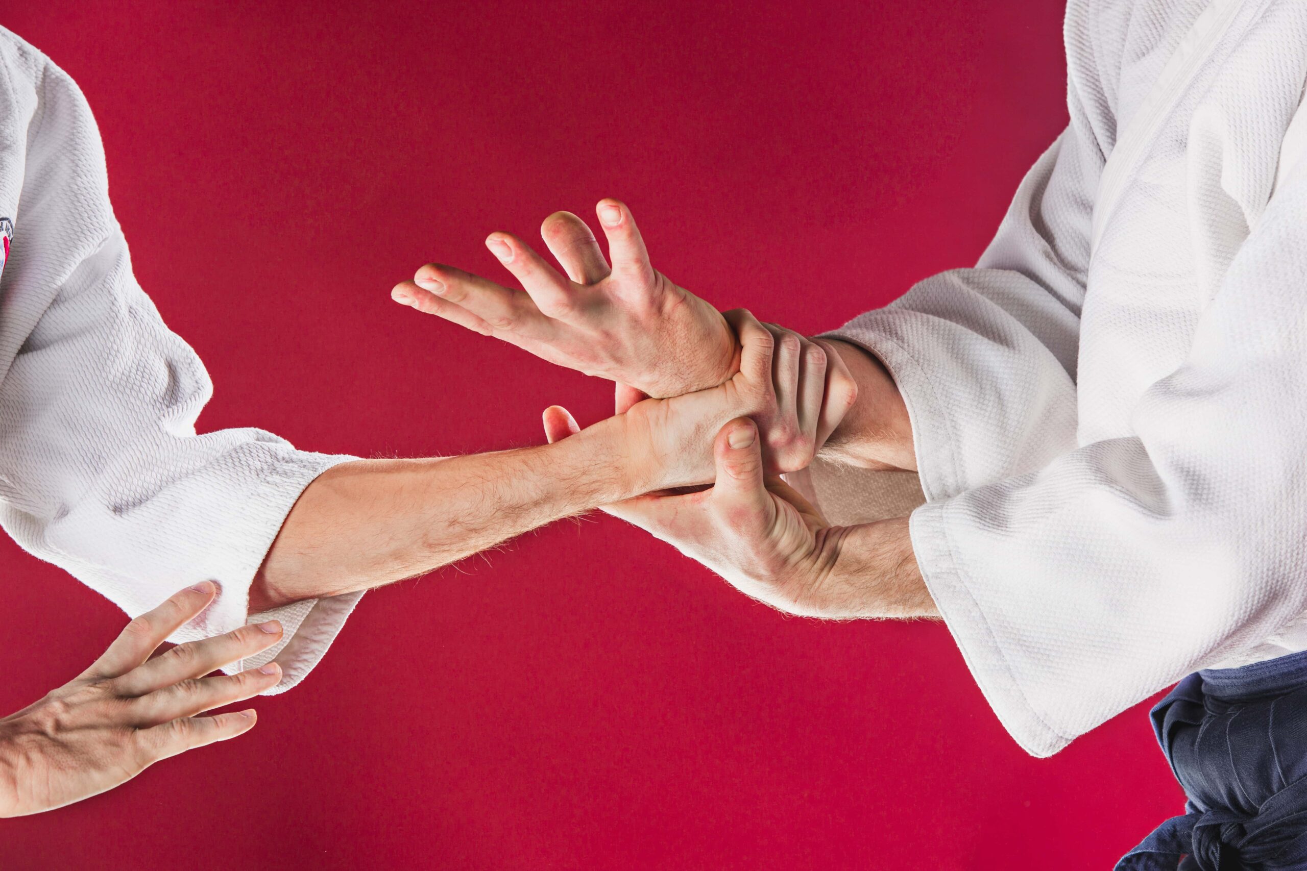
UFC fighter Tony “El Cucuy” Ferguson is widely regarded as one of the best lightweights in the history of the UFC.
Tony’s left ulna bone was fractured while fighting against Michael Johnson in a 2012 UFC event. El Cucuy’s forearm injury has healed completely and he’s fought many times since his treatment.
Forearm Injury Causes
Martial arts and combat sports utilize the arm for defensive moves such as blocking strikes and kicks, throws, offensive moves such as armbars, grappling, hammer blows and punches, so it’s no surprise that the arm is the most commonly injured body part in fighters.
Forearm fractures and injuries are common in mixed martial arts. The natural reflex of defense is to use the forearm. In many combat sports, the forearm is used for grip. And grip strength is used to control a wrist or holding a submission. Grip strength can win or lose a fight. While using the forearm to block a forceful kick, the bony part is exposed. The bony part tends to be the ulna. In most forearm injuries, it’s this ulna that is injured.
Forearm Injury Symptoms



Types of Forearm Injuries
There are various fractures involving the bones in the forearm. These include Galeazzi fractures where the radius is broken independently. Monteggia fractures involve both the radius and ulna. And then there’s’ the nightstick fracture which is most common in mixed martial arts where the ulna is fractured independently.
Arm Injuries Due to Hand to Hand Combat Training Are Common
Learn When to Get Help Versus Training Through the Pain
Related Injuries
This occurs when the bones of the forearm have been heavily impacted resulting in the hairline or non-displaced fractures, or comminution fractures either with or without multiple fragments. This injury is sometimes called the “nightstick” injury since it traditionally resulted from protecting oneself from attackers using a club, heavy bar, or nightstick.
Symptoms
The symptoms will be obvious as there will be extreme pain after a training accident, attack, or fight event. Symptoms may include:
Causes
Forearm fractures in martial arts combat events most often occur when a fighter is using the arm to block a kick to the head or shoulder resulting in a bone fracture to the outer forearm.
To learn how Nightstick Fracture is diagnosed, Click Here
The Galeazzi fractures account for about 7% of all fractures in adults. This is a forearm fracture common in other sports like football and wrestling too. This is a fracture of the middle to distal one-third of the radius. It is often associated with either the dislocation or subluxation of the distal radioulnar joint.
Symptoms
Causes
The radius and ulna in the forearm are stabilized by three ligaments. The interosseous membrane disperses the axial load to the forearm. This distribution is about 60% to the radiocapitellar joint and 40% to the ulnohumeral joint.
When a fighter falls on an outstretched hand with an extended wrist and a hyperpronated forearm, the load is displaced. All the energy from the radial fracture now gets transmitted to the ulnohumeral joint. This dislocates the distal radioulnar joint. In MMA, falls on an outstretched arm are not as common as in other sports, but accidental injuries can cause Galeazzi fractures.
To learn how Galeazzi Fracture is diagnosed, Click here.
In a Monteggia fracture, the proximal ulna if fractures along with a dislocated radial head. They are commonly seen in athletes who sustain falls or receive a direct blow to the forearm. This usually occurs while the elbow is extended and the forearm is hyperpronated.
The fracture is caused when the energy from the ulnar fracture is transmitted along the interosseous membrane. It causes the proximal quadrate and annular ligaments to rupture. This disrupts the radiocapitellar joint. These are not as common with them accounting for about 1% of all forearm fractures.
The Monteggia fractures are classified into four categories
To Learn How Monteggia Fracture are Diagnosed, Click Here
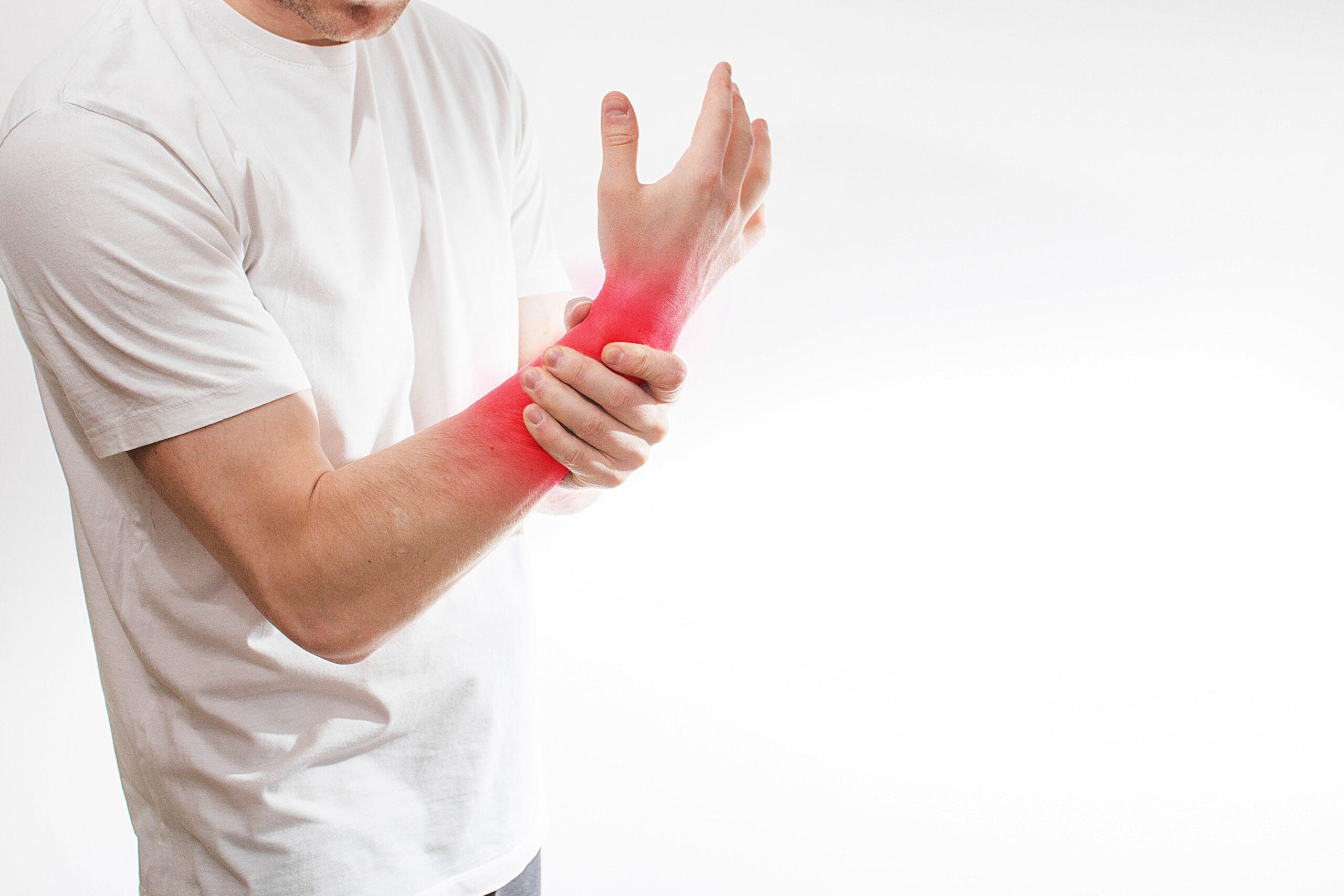


Common Forearm Injuries
For common forearm injuries like sprains and strains, refer to our Common Injuries section.
Forearm Injury Diagnosis
Forearm fractures are complex fractures. The Monteggia fracture is the most difficult to diagnose. If forearm fractures are missed, the morbidity is very high. It can result in permanent disability, limited motion, and chronic pain. Nerve injuries also persist not from the injury but the forceful reduction of the bones as well. Galeazzi fracture typically also have a high rate of morbidity. They are easily missed on radiographs. Muscle entrapment may also complicate the diagnosis. It is imperative that when forearm fractures are suspected images are done right away.


MMA fighter Ryan Jimmo told officials during his bout that he could feel the bone moving in his forearm. Jimmo had a nightstick fracture where luckily for him the ulna had a clean break. Officials failed to take him seriously until he was crying in pain. Not all fractures break the skin and so closed fractures can also occur.
Injury Specific Diagnosis
Physical Exam
On physical examination, a thorough examination of the skin is done to rule out any open fractures. These require immediate surgical care. Next, a thorough examination of the elbow and wrist is done. This is to rule out any associated Monteggia fracture or other wrist injuries. Doctors will test the range of motion at the elbow. They will palpate the area for any "clicks" or "clunks." This is done in particular at the site of the radial head. They will also do a "shuck test" at the wrist. This is just to check for weakness or instability of the distal radioulnar joint (DRUJ).
Imaging
X-ray films are the most important test. X-rays of the elbow with a lateral view is done. This is to check if there is an appropriate alignment of the radial head. Any misalignment will cast suspicion as to a Monteggia fracture. X-rays of both wrists are done to assess the wrist joint. The films are also done during pronation and supination bilaterally. CT scans are not necessary. Advanced imaging like CT or MRI is used to rule out other diagnoses.
Lab Tests
Polytrauma fighters and those scheduled to undergo surgeries will require tests. These include blood tests, chest Xray and an ECG. Blood tests will include hemoglobin, BUN/Creatinine, Glucose, PT, PTT, SGOT, ALK, CMP, and others. In females, a pregnancy test will also be required.
To learn how Nightstick Fracture is treated, Click Here
Physical Exam
On physical exam, the orthopedic doctor visually inspects the skin and soft tissue. They will look for visible bony deformities, lacerations on the skin where the bone might protrude, muscle contusions, damage to the and neurovascular deficits. The open fractures will necessitate immediate surgical intervention without a further physical exam.
They will gently palpate the bone to identify deformities and point tenderness. The stability of the proximal and distal joints is assessed. This will help to identify any simultaneous injuries. A detailed neurovascular exam is performed to look for signs of acute compartment syndrome. They will examine the median and radial nerve distribution to see if there is any nerve damage.
Imaging
X-ray films are warranted. Usually, an anteroposterior and lateral view is used to identify the fracture. An oblique view is ordered to classify the injury. Classifying the injury will influence the treatment. X-ray films are obtained from the distal wrist and proximal. This is to rule out any concomitant injury. On Xray imaging, signs of disruption of the distal radioulnar joint (DRUJ) are seen as widened DRUJ, an ulnar styloid fracture, dorsally displaced ulna on the lateral view, and shortening of radius more than greater than 5 mm. In all cases of forearm fractures, Xray films of the normal limb are taken for comparison. Usually, Xray films are enough. If fractures involve non-union, then, a CT scan and magnetic resonance imaging (MRI) can help evaluate for ligament tears and disruption.
Lab Tests
If surgical treatment is required, then it will need blood tests and an ECG. Blood tests will include hemoglobin, BUN/Creatinine, Glucose, PT, PTT, SGOT, ALK Phos. In females, a pregnancy test will also be required.
To Learn how Galeazzi Fracture is treaded, Click Here
Doctors will visually inspect the skin and soft tissue for visible bony deformities, contusions of the muscles, skin lacerations, damage to ligaments and tendons and neurovascular deficits. They will palpate the bones and identify tenderness along the injured bone. They will examine both, the proximal and distal joint. This is to rule out any coexisting injuries. They will look for signs of acute compartment syndrome and radiating pain. The examination of the radial and median nerves is important for identifying nerve damage.
Xrays films are essential for all forearm fractures. To identify the fracture, an anteroposterior and lateral view is done. An oblique view can classify the injury. X-ray films of the distal wrist and proximal elbow are also done. Advanced imaging is not needed. CT and MRI are done to assess tears and disruption in the stabilizing ligaments.
Pre-operative tests include blood tests, chest Xray and an ECG. Blood tests will include hemoglobin, BUN/Creatinine, Glucose, PT, PTT, SGOT, ALK, CMP, and others. In females, a pregnancy test will also be required.
To learn how Monteggia Fracture are treated, Click Here


Diagnoses of Common Forearm Injuries
The Common Diagnoses section will discuss the X-rays and other radiological modalities to diagnose skeletal fractures.
Forearm Injury Treatment
The treatment of the forearm fractures depends on the type of fracture and the joints involved. Fighters who suspect forearm fractures must see an orthopedic doctor. The length of treatment depends on multiple factors. Some of these include the severity of the injury, age, the type of injury, and also the intended use after the injury. Typically fractures take about 6 weeks to heal through remodeling.
If surgery is warranted, the rehabilitation takes place only 8 weeks after the surgery. The goal of any post-surgical rehab is to help athletes gain a full range of motion and fine motor skills. A good sign of healing is when pain is absent and return to full motion. Fighters who will return to high demand activity require 12-16 weeks of rehabilitation care. The surgical hardware like screws, pins, and plates are usually permanent in the forearm and are in place for life.
Injury Specific Treatment
Emergency Treatment
Historically many of these fractures are closed and hence after ice and splinting, they should be seen by an orthopedic doctor. Pain medications are given to relieve the pain.
Medical Treatment
Non-displaced fractures may be treated with a long-arm cast. A long arm cast is used to completely immobilize the fracture and allow healing. The cast usually remains for 6-8 weeks at which time a removable fracture brace may be used until completely healed. Displaced and/or comminuted fractures usually require surgery to insert a metal plate. This surgery usually allows the new solid bone to grow in the gap. This plate may or may not be removed once healing is achieved. Fighters will often decide to leave the plate in place as it gives more strength to the area, and removal means more downtime before resuming fighting.
Home Treatment
For nightstick fractures, early mobilization has proven helpful for recovery. To promote the union of the bone, doctors advise against prolonged immobilization and functional bracing. Below elbow, bracing is superior to above elbow bracing.
Emergency Treatment
These fractures require an orthopedic consult. Rest, ice, immobilization, and elevation is the first step for a suspected fracture. Fighters should have their forearms placed in a sugar tong splint. This fracture will require surgery eventually.
Medical Treatment
Surgery is always preferred since closed reduction adults tend to have a poor prognosis. First, the radial shaft fracture is fixed with rigid fixation. Then the stability of the DRUJ is assessed intra-operatively. If the joint is stable, then it is pinned with two K wires. If the joint is unstable, the ligament is repaired after which it is pinned. If there is an accompanying ulnar styloid fracture, then the ligament is repaired, and the styloid is fixed using a screw lag or tension band wire.
Home Treatment
At home, rest is advised. The forearm is cast in supination in an elbow splint. The fighter must be careful and watch for signs of wound infection and compartment syndrome. These signs include crepitus, warm muscles, tense, shiny skin, and pain.
All Monteggia fractures are considered highly unstable and will require surgical intervention. Immediate, orthopedic consult is warranted. Rest, ice, immobilization, and elevation are the first steps after a fracture is suspected. Fighters must place their forearms in a sugar-tong splint and advised urgent referral to an orthopedic doctor.
Despite the closed reduction, Monteggia fractures in adults are more prone to angulation and shortening. Therefore, these fractures are fixed by a procedure called open reduction internal fixation (ORIF). A compression plate is fixed with six cortical screws proximally and distally. Once the plate is in place, the radial head dislocation is easily reduced.
After surgery, the splints and angles of casting all depend on the type of fracture. The forearm is placed in a long-arm splint. It is fully supinated with elbow flexion around 100-degrees for Types 1, 3, and 4 fractures. In the case of type 2 fractures, the elbow is splinted at 70-degrees. Pain medications are started and rehab begins 2 weeks after surgery. Despite this, fighters still complain of chronic pain and limited motion.
